Bioimpedance
How do BIA and BIS evaluate body composition?
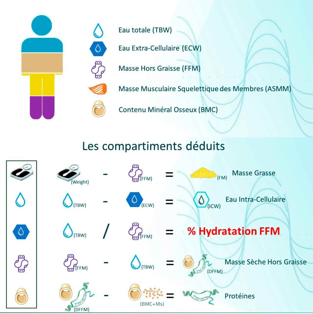
Body composition corresponds to the distribution between fat mass, skeletal muscle mass, and water in the body, providing a much more relevant evaluation than weight alone or BMI.
Bioimpedance (Bioelectrical Impedance Analysis, BIA) is a non-invasive and painless method for analyzing these compartments. It relies on how body tissues react to an alternating electrical signal of very low intensity, imperceptible to the individual.
This response depends notably on tissue hydration and on the properties of cell membranes. By integrating these parameters into validated prediction models, BIA allows for the estimation of fat-free mass and its associated compartments, notably skeletal muscle mass and total water.
Bioelectrical Impedance Spectroscopy (BIS) goes even further: it measures the body’s electrical response over a wide spectrum of frequencies, allowing for a finer characterization of fluid compartments and cellular properties (such as membrane capacitance and characteristic frequency). This spectroscopic approach improves the physiological robustness of the model and provides direct quality control thanks to the analysis of signal coherence across the entire spectrum.
Thanks to their simplicity of use and the speed of obtaining results, BIA and BIS provide essential information on nutritional and hydration status, constituting reliable tools for clinical follow-up.
BIA is strongly correlated with total body water, which allows for the precise estimation of fat-free mass using predictive equations. However, since adipose tissue is very poorly linked to the measured parameters, its direct estimation is less reliable. Thus, there is a scientific consensus to calculate fat mass by difference, by subtracting fat-free mass from total weight, in accordance with the body compartment model.
Parameters derived from the bioelectrical signal
Beyond the estimates derived from body composition models, certain parameters are calculated directly from the measured electrical signal. These so-called “raw” indicators do not rely on predictive equations but reflect the intrinsic electrical properties of biological tissues.
Phase Angle (PhA)
The phase angle is derived from the relationship between resistance (R) and reactance (Xc). It reflects the capacitive behavior of cell membranes and serves as a global indicator of membrane integrity and active cell mass.
Physiologically, the PhA reflects the capacity of cells to maintain their structure, intracellular hydration, and functionality. A decrease in the phase angle can be associated with tissue alteration, fluid imbalance, or a catabolic state. Monitoring it over time provides valuable qualitative information on the evolution of cellular health.
Impedance Ratio (IR)
The impedance ratio (IR) is generally expressed as the ratio between the impedance measured at high frequency and that measured at low frequency. It provides complementary information on the distribution of body fluids.
The IR makes it possible to assess the balance between intra- and extracellular compartments as well as the structural coherence of tissues. An elevation in the IR may reflect a change in membrane permeability or an alteration in fluid distribution.
The joint interpretation of the PhA and IR reinforces the physiological robustness of the analysis. These parameters enrich the clinical assessment by providing a qualitative reading of the biological tissue, beyond the simple quantitative estimation of fat or muscle mass.
Factors influencing the accuracy of BIA measurements
The accuracy of a BIA measurement depends on several technical and physiological parameters. Among the most determining are the quality of contact between the electrodes and the skin, the body position during the measurement, and the connection mode used by the device.
Contact points
Hand-foot analysis for a consistent whole body measurement.
Spectroscopy
54 measurement points, instant quality control.
Multi-algorithms
No simplified deduction: each compartment is calculated independently.
Clinical reference
Tetrapolar method and algorithms validated by literature.
Importance of contact points
The contact points determine the signal path in the body and therefore the compartments actually traversed. Depending on the combination used (hand-hand, hand-foot, foot-foot), the measurement provides information on a different body volume.
Foot - Foot
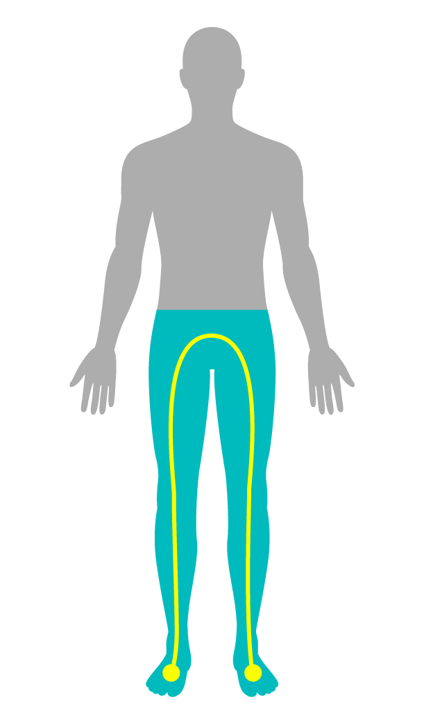
- Measurement limited to the lower body
- Trunk and upper body estimated, not measured
- Variable accuracy depending on sex and morphotype
Hand - Foot
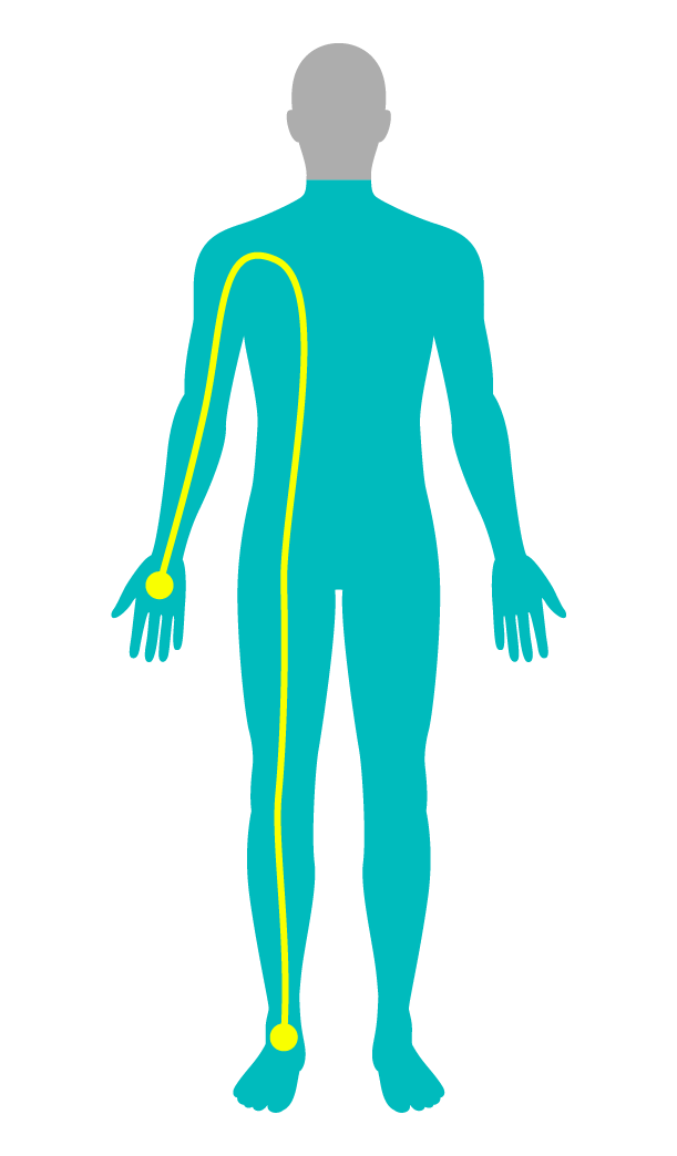
- Measures the whole body: limbs + trunk
- Complete and more reliable analysis
- Preferred method in clinical practice
Hand - Hand
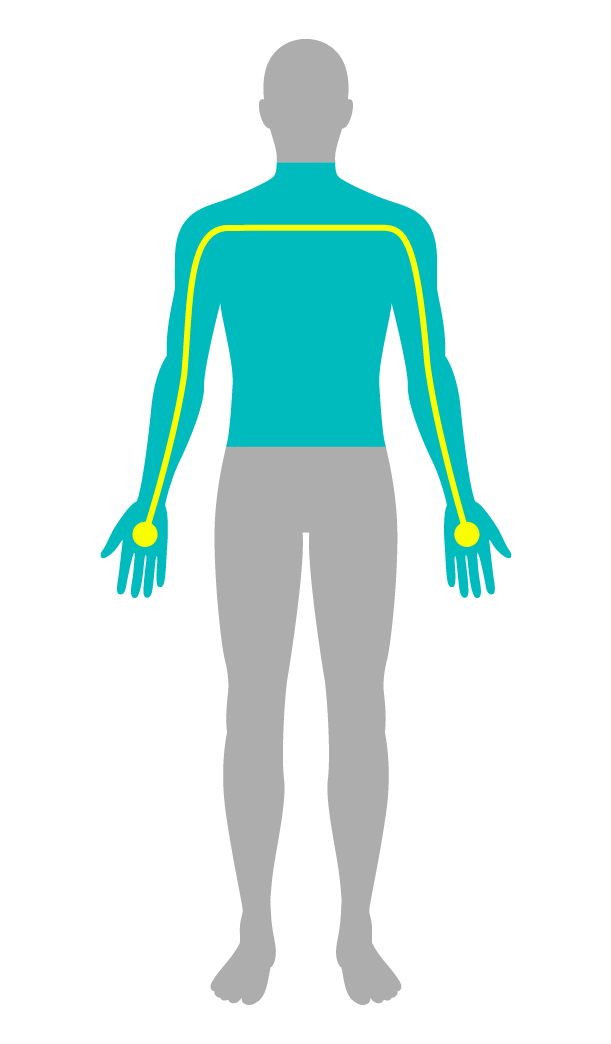
- Measurement limited to the upper body
- The lower body is estimated, not measured
- Accuracy dependent on morphotype and sex
Aminogram devices rely on the hand-foot measurement, the reference method for complete and reliable body analysis, recommended by ESPEN and the Haute Autorité de Santé.
Measurement positions
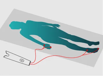
The lying position
Homogeneous distribution of fluids after 15–20 min
- Excellent precision
- Longer stabilization time
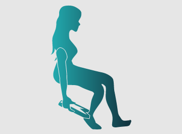
The sitting position
Stable, fast, and reproducible position
- Practical in consultation
- Comfortable for the patient
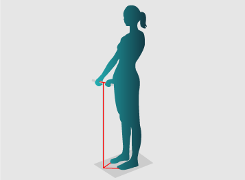
The standing position
Distribution of fluids towards the lower body
- Quick to implement
- Poorer water homogeneity
Aminogram devices allow for measurement in the sitting or lying position, with or without cable, in order to combine simplicity of use and clinical precision.
The sitting position, easy to implement, is perfectly adapted to routine consultations. The lying position, on the other hand, offers an ideal environment for the most fragile populations and for evaluations requiring optimal fluid stabilization.
Mesure directe et indirecte
Pour réaliser une mesure, deux modes de connexion peuvent être utilisés :
Mesure indirecte
(avec câbles)
- : positionnement flexible, compatible avec les mesures assise ou allongée.
- : la résistance des câbles peut légèrement influencer la mesure.
Mesure directe
(sans câbles)
- : installation rapide, mesures stables non affectées par les câbles, aucun consommable.
Les dispositifs Aminogram permettent d’utiliser la mesure directe ou indirecte selon le contexte. La mesure directe offre rapidité et reproductibilité, tandis que la mesure indirecte est adaptée aux mesures assises ou allongées. Cette flexibilité garantit une utilisation précise et confortable, quel que soit le patient ou l’environnement.
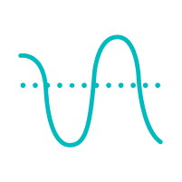
Single-frequency, multi-frequency and spectroscopy
BIA measures the electrical response of tissues, influenced by hydration and cell membranes. This response varies according to the frequency of the current:
- Low frequencies (< 7 kHz) : the current remains in the extracellular medium.
- High frequencies (> 50 kHz) : it crosses the membranes and accesses intracellular water.
It is this variation that allows distinguishing fluid compartments and improving analysis precision.
Single-frequency
- Single frequency (50 kHz)
- Measures total impedance and phase angle
- Does not fully cross the cell membrane
- Estimated fluid compartments → limited precision
Multi-frequency
- Multiple frequencies (1–1000 kHz)
- Differentiates intra- / extracellular water
- Real data on fluid compartments
- More complete models (up to 6 compartments)
Spectroscopy

- Continuous spectrum scanning (1–1000 kHz, 54 points)
- Fine analysis of tissue electrical response
- Access to physiological parameters (capacitance, characteristic frequency)
- Instant quality control (Cole-Cole curve)
Spectroscopy, based on the Cole-Cole model, offers a finer reading of cellular properties and real-time quality control, ensuring the reliability and consistency of each measurement.

Single-algorithm vs Multi-algorithms
The electrical parameters measured in BIA (impedance, resistance, reactance, phase angle) must be interpreted using algorithms. Two approaches coexist:
Single-algorithm
(deductive)
Relies on a unique model assuming constant body hydration (e.g. 73.3% of fat-free mass), identical for all.
The compartments are deduced from fixed coefficients.
Example:
- Fat-free mass = Total water × 0.733
- Proteins = Fat-free mass × 0.198
Fast method but sensitive to physiological variations (hydration, pathologies, morphotypes).
Multi-algorithms
(non-deductive)
Uses several independent models, each dedicated to a body compartment (total water, fat-free mass, fat mass, body cell mass, etc.).
Each compartment is calculated according to a specific algorithm derived from the measured bioelectrical properties.
Allows for determining the real hydration level of the fat-free mass, without arbitrary assumptions.
More consistent and precise analysis, adapted to individualized clinical monitoring.
All Aminogram devices rely on a Multi-algorithm approach, ensuring physiologically consistent estimates, better clinical precision, and compatibility with prediction equations validated in scientific literature.
Tetrapolar measurement: the reference validated by ESPEN
Tetrapolar measurement uses four distinct electrodes: two to inject current and two to measure voltage. This separation of circuits ensures a precise, stable, and reproducible measurement, without influence from skin contact.
Unlike octopolar systems, which artificially segment the body and multiply sources of error, tetrapolar measurement provides a global reading, consistent with human physiology.
Recommended by ESPEN and the Haute Autorité de Santé, it constitutes the international reference method — and the basis of all Aminogram devices.
Performance complying with scientific requirements (Gold Standard)
Our design approach relies on published data in order to guarantee accuracy consistent with reference methods. The algorithms integrated into our devices are derived from models validated in scientific literature, ensuring a reliable estimation of the various body compartments.
We have also conducted clinical evaluations confirming the accuracy of measurements for fat-free mass, fat mass, appendicular skeletal muscle mass, and bone mineral content. All results and methodological details are presented in our scientific booklet.